-
NATIONAL

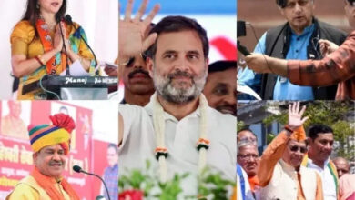
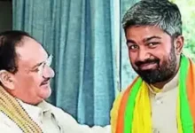
Lok Sabha Elections 2024: Omar and Mehbooba request that the…
On Friday, PDP president Mehbooba Mufti and NC leader Omar Abdullah urged the Election Commission…
Read More » -
-
-
-


-
UP STATE
Important Spectators in Phase 2 of the Lok Sabha Election…
The second round of India’s parliamentary elections, which will take place in 13 states and…
Read More » -
-
-
-
BIHAR
When it comes to soil health and fertility schemes, Bihar…
Patna: At a ceremony conducted in New Delhi on Thursday, Bihar was given a certificate…
Read More » -
-
-

-
ENTERTAINMENT
Kajol pushes followers to guess the specifics of her fitness…
Bollywood superstar Kajol recently shocked fans by sharing an amusing glimpse into her exercise regimen.…
Read More » -
ENTERTAINMENT
The European Program of Hot Docs Examines Work Conditions, Right-Wing…
The sixth iteration of Toronto’s Hot Docs Film Festival’s The Changing Face of Europe segment…
Read More » -
ENTERTAINMENT
Chinese Internet Films Reimagined as PopC Linear Channel – World…
With material provided by mainland China streamer iQiyi, Celestial Tiger Entertainment, the operator of Asian…
Read More » -
ENTERTAINMENT
Kerry Condon on Drawing Inspiration from “Trainspotting” for the Upcoming…
Kerry Condon is returning to her home country of Ireland for her next film role.…
Read More »


-
INTERNATIONAL
Is a UFO sighting in New York? Unidentified “flying cylinder”…
New York: An aircraft passenger saw a weird “flying cylinder” above New York, which baffled…
Read More » -
-
-
-


-
HEALTH
High risk of childhood stunting is associated with hilly terrain,…
Research published on Friday suggests that children under the age of five who live at…
Read More » -
-
-


-
LIFESTYLE

Discover The Benefits of Adventurers Traveling to Rishikesh
Why Is Rishikesh Well-Known? Located at the base of the Himalayas, Rishikesh is revered as…
Read More » -
-

-

-
SPORTS
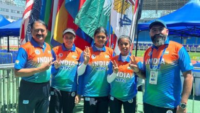
at the compound mixed team final, Jyothi Surekha Vennam and…
In the current Archery World Cup, Jyothi Surekha Vennam and Abhishek Verma’s compound mixed team…
Read More » -
SPORTS
Indian ladies want to excel in the Uber Cup, while…
Star singles players will want to have a steady run as the Indian men begin…
Read More » -
SPORTS
Ten Hag was ecstatic with United’s comeback victory
After inspiring a tense 4-2 victory against Sheffield United on Wednesday, Bruno Fernandes relieved some…
Read More » -
SPORTS
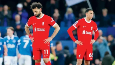
“I only have regrets,”
After Liverpool’s Premier League championship campaign was severely damaged by a 0-2 setback at Everton…
Read More » -
SPORTS
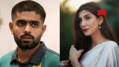
‘Rude Reply’ On Marrying Babar Azam: Actor Nazish Jahangir of…
Actor Nazish Jahangir of Pakistan allegedly received backlash from Babar Azam’s followers on social media…
Read More »